MICCAI 2012 Challenge on
Multimodal Brain Tumor Segmentation
1 October 2012 - Acropolis Convention Center - Nice, France
| New: A pre-print of the manuscript The Multimodal Brain Tumor Image Segmentation Benchmark (BRATS) is available - please cite this to refer to the BRATS challenge in your papers! |
Background and set-up
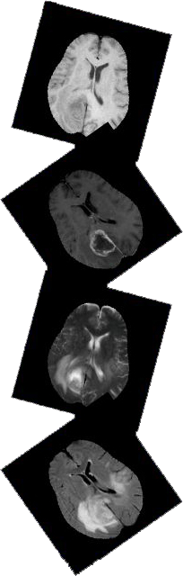
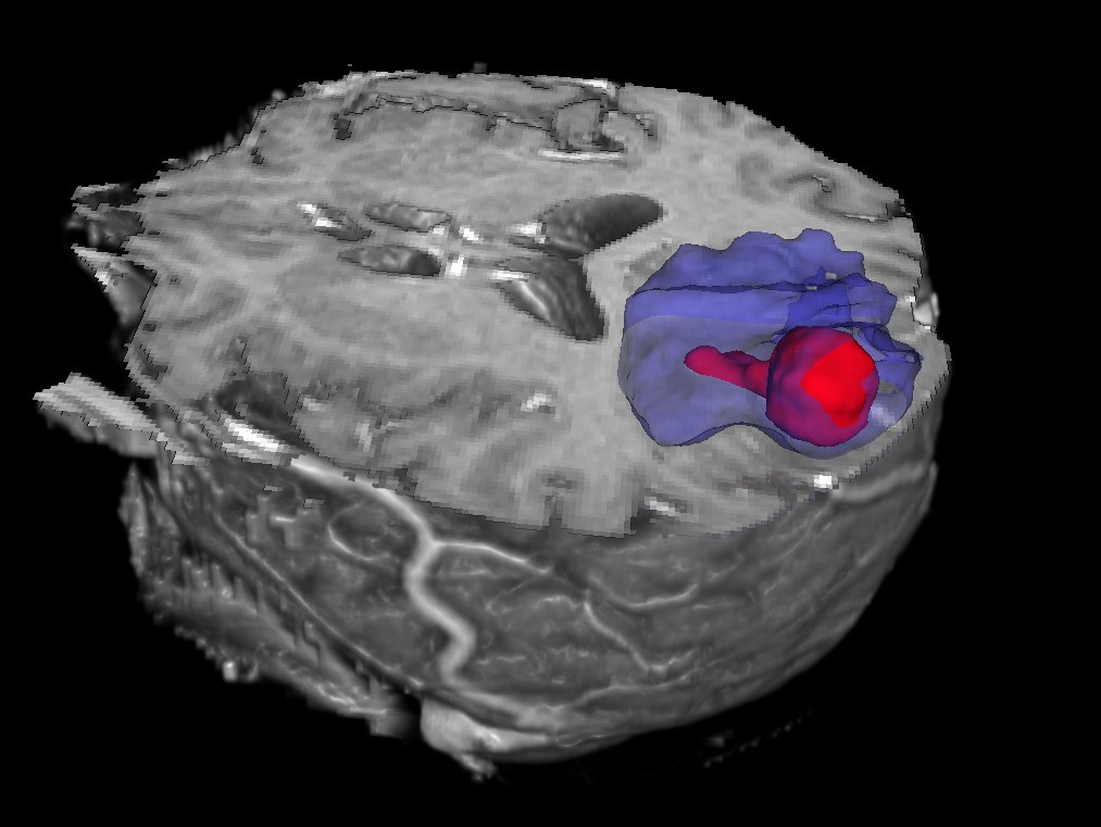
Because of their unpredictable appearance and shape, segmenting brain tumors from multi-modal imaging data is one of the most challenging tasks in medical image analysis. Although many different segmentation strategies have been proposed in the literature, it is hard to compare existing methods because the validation datasets that are used differ widely in terms of input data (structural MR contrasts; perfusion or diffusion data; ...), the type of lesion (primary or secondary tumors; solid or infiltratively growing), and the state of the disease (pre- or post-treatment).
In order to gauge the current state-of-the-art in automated brain tumor segmentation and compare between different methods, we are organizing a Multimodal Brain Tumor Segmentation (BRATS) challenge in conjunction with the MICCAI 2012 conference. For this purpose, we are making available a large dataset of brain tumor MR scans in which the tumor and edema regions have been manually delineated. In addition, we also provide realistically generated synthetic brain tumor datasets for which the ground truth segmentation is known.
Challenge format
Teams wishing to participate in the challenge should download the training data for algorithmic tweaking and tuning. The teams should then evaluate their segmentation performance on the training data, and submit a short paper describing the results and the segmentation method that was used. On the day of the challenge itself, an independent set of test scans will be made available and analyzed on the spot by each team, after which the methods will be ranked according to their performance. The challenge day will conclude with a round-table discussion of the obtained results as well as invited talks by clinical experts.
In the weeks following the challenge participating teams will be invited to contribute to a joint paper describing and summarizing the challenge outcome, which we will then submit to a high-impact journal in the field.
A FAQ with more details can be found here.